Wednesday, August 6, 2014

Other Pulmonary Diseases

Other Pulmonary Diseases-

-Acute Respiratory Distress Syndrome-


-Acute Respiratory Distress Syndrome (ARDS) is a distinct type of hypoxemic respiratory failure

-When healthy lungs are disrupted by lung injury, infection, of fluid, there is a disruption of surfactant causing excess fluid in both the interstitial spaces and the alveoli

-The alveolar injury produces alveolar damage and causes release of inflammatory cytokines such as TNF and interleukins

-Damage of the capillary endothelium allows protein to escape from the vascular space.  The fluid then pours into the interstitial spaces and overwhelms the lymphatics

-As a result of this there is impairment of gas exchange, decreased lung compliance, and increase pulmonary artery pressure

-Possible etiologies of ARDS include:  sepsis, aspiration, pneumonia, trauma, burns, multiple blood transfusions, pancreatitis, drug overdose, near drowning, pulmonary contusion, smoke inhalation, CABG, pulmonary contusion, venous air embolism, drug reaction, amniotic fluid embolism, pulmonary edema, and BOOP

-Signs and symptoms of ARDS typically appear within 6-72 hours of the initial insult or injury and decline rapidly

-Patients typically present dyspnea, cyanosis, hypoxemia, diffuse crackles, tachycardia, diaphoresis, and accessory muscle use.  Patients are typically coughing and will have chest pain

-Patients typically are hypoxemic and a wide A-a gradient

-High FiO2 is needed to get desired oxygenation

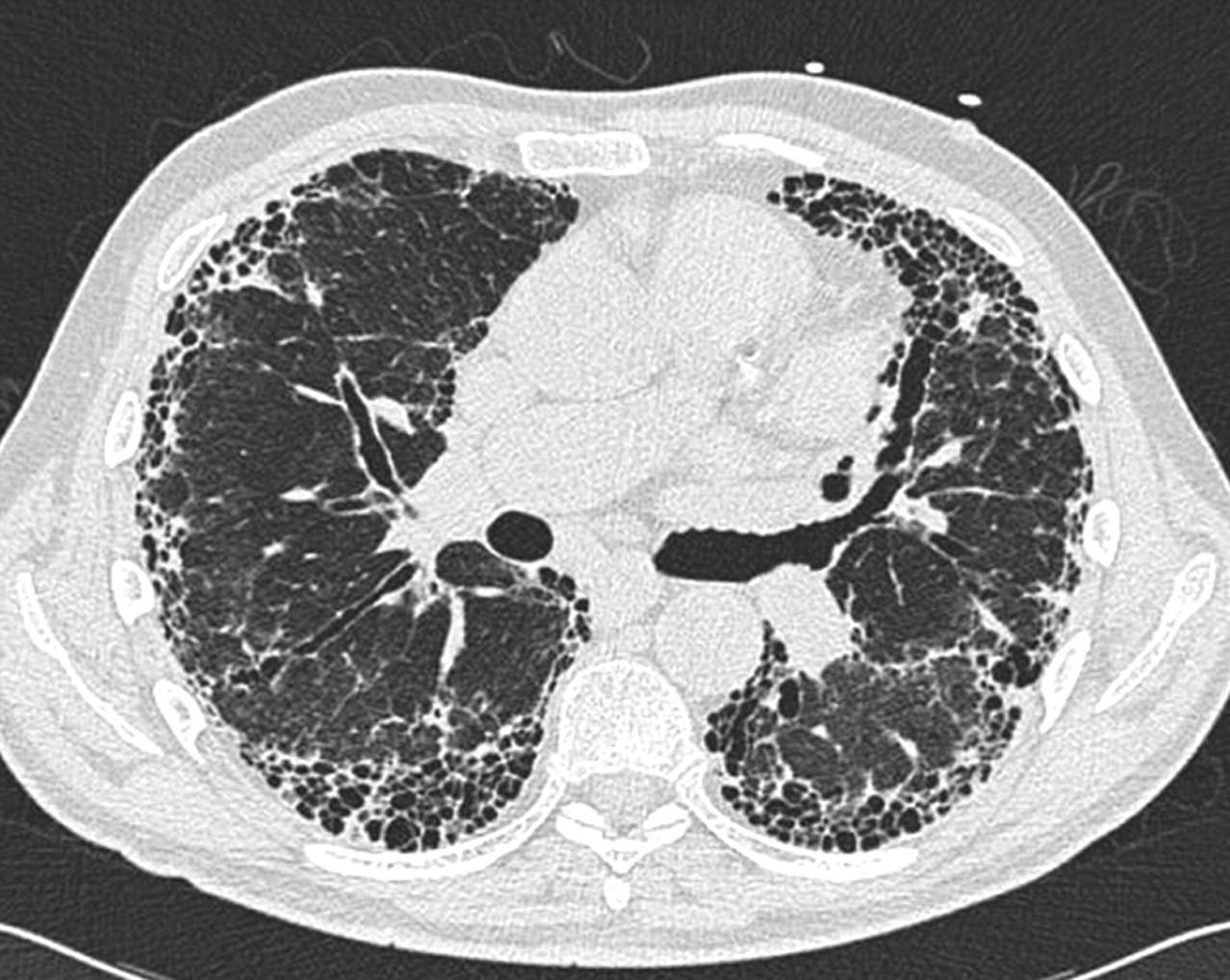
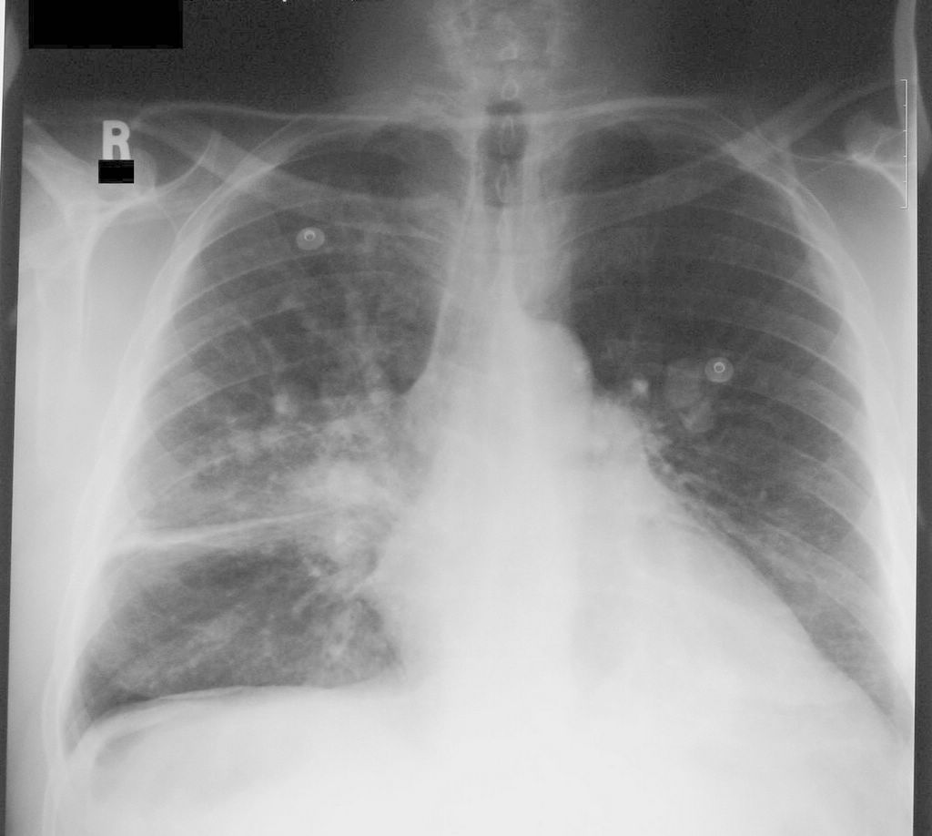
-Chest X Ray typically shows bilateral alveolar infiltrates, and CT scan will show widespread patchy airspace opacities that are more apparent in dependent lung zones.  This will likely progress to a ground glass appearance

-Most patients require moderate to high concentration of inspired oxygen over the first several days.  The patients who survive will have better oxygenation and decrease in alveolar infiltrates which will allow ventilator weaning to begin

-Complications of ARDS are related to mechanical ventilation:  barotrauma, nosocomial pneumonia. May also have delirium, GI bleed due to stress, thrombosis, and catheter related infections

-Once the initial injury or insult is past or treated, the patients who respond can be weaned from the ventilator and FiO2 can be weaned


-Hyaline Membrane Disease-


-Hyaline membrane disease (HMD) is also known as Respiratory Distress Syndrome (RDS) or Infant Respiratory Distress Syndrome (IRDS)

-The pathophysiology behind HMD is that there is a deficiency of surfactant, usually due to prematurity, that reduces the alveolar surface tension, which decreases the pressure needed to keep the alveoli open, and maintain alveolar stability

-When the surfactant is deficient, the infant cannot generated the increased inspiratory pressure to keep the alveoli open, and results in alveolar collapse and diffuse atelectasis

-This subsequently leads to decreased lung capacity and low functional residual capacity.  Hypoxemia then results.  This may lead to right to left shunting and cause a further VQ mismatch

-Prior to exogenous surfactant administration, uncomplicated HMD typically progressed for 48-72 hours until there was production of endogenous surfactant

-Now with treatment with exogenous surfactant (Survanta), there is dramatic improvement of pulmonary function, and shortening of clinical course.  Use of CPAP has improved the clinical course of HMD


-Foreign Body Aspiration-


-Foreign body aspiration is can be potentially a life threatening event

-Foreign bodies are most likely to be aspirated into the right side secondary to the angle of the right mainstem bronchus

-Inorganic material such as glass or metal may cause little tissue inflammation but can result in direct airway injury if they are sharp

-Organic material such as nuts can cause significant inflammation and granulation tissue formation resulting in stenosis

-Aspiration of medications such as iron tablets in pill form can cause airway inflammation and ulceration

-X-Ray may confirm the presence of foreign body; however, direct visualization with bronchoscopy is need for definitive diagnosis and removal.

-In cases of life threatening aspiration initial support should focus on treating the airway and removing the obstruction.  If it is above the larynx may be able to be removed with forceps, if below the larynx will need bronchoscopy for removal.


Restrictive Pulmonary Disease

Restrictive Pulmonary Disease-

-Idiopathic Pulmonary Fibrosis-


-Idiopathic pulmonary fibrosis (IPF) is a chronic progressive disorder of the lower respiratory tract that typically affects adults over the age of 40

-The pathogenies of IPF is unknown.  Certain risk facts include cigarette smoking, viral infection, environmental pollutants, chronic aspiration, genetic predisposition, and drugs

-Patients present with progressively worsening shortness of breath and eventually hypoxemia

-The severity of the disease is determined by pulmonary function testing

-Pulmonary function test usually demonstrate a reduction of the FVC and DLCO

-Advance disease is characterized clinically by dyspnea on mild exertion (less than 300 feet) and a requirement for supplemental oxygen at rest or exertion.  Extensive honeycombing is seen on imagine studies such as chest x ray or CT scan of chest

-The prognosis of IPF is poor.  Only 20-30 percent of all patients will survive greater than 5 years

-Elements of supportive care for IPF include eduction, supplemental oxygen if needed, pulmonary rehab, and vaccines against influenza and Streptococcus pneumoniae

-No medications have been show to slow lung damage

-Potential medication therapies include pirfenidone and phosphodiesterase inhibitors

-Lung transplantation is an option if patients with IPF meet certain conditions


-Pneumoconiosis-


-Coal workers pneumoconiosis comes from inhalation and deposition of silica free coal dust particles that induce the formation of coal macules in the alveoli

-Findings with pneumoconiosis give pigment and reticulin fibers that accumulate in the peri-hilar location.  Over time black amorphous masses develop and liquify in the center.  There is cavitation that results in ischemic necrosis

-On x ray you see rounded small nodular opacities less than 1 cm usually in the upper lobes.  Eventually there is a confluence of large opacities that lead to massive progressive fibrosis

-Findings of PFT's are similar to IPF.  There is a reduced FVC and DLCO.

-May have some improvement when the offending agents are removed

-Much like IPF, treatment is largely supportive


-Sarcoidosis-


-Sarcoidosis is a multisystem granulomatous disorder of unknown etiology and is characterized by non-caseating granulomas in involved organs

-Sarcoidosis typically presents in young adults with pulmonary reticular opacities, bilateral hilar adenopathy, and skin, joint, or eye lesions

-The exact etiology and pathogenesis of sarcoidosis are unknown

-Sarcoidosis most often involves the lung (70 percent of the time)

-Diffuse interstitial lung disease is the classic type of lung development

-Common presenting symptoms of sarcoidosis are cough, dyspnea, chest pain, fatigue, malaise, fever, and weight loss

-Patients with lung parenchyma sarcoidosis typically will have crackles heard on lung exam.  Wheezing is heard when there is endobronchial involvement or traction bronchiectasis due to scarring

-Children with sarcoidosis usually do not become symptomatic

-Initial workup for suspected sarcoidosis patient should include:  CBC, BMP, tuberculin skin test, PA and lateral chest x ray, pulmonary function testing, EKG, and eye exam by an ophthalmologist

-Diagnosis is made my biopsy of involved organ.  Can do skin biopsy, or lung biopsy to confirm most commonly

-The majority of patients with pulmonary sarcoidosis have non progressive disease or experience spontaneous remission

-Oral glucocorticoids have been used for the relief of symptoms and control of potentially disabling respiratory impairment


Tuesday, August 5, 2014

Pulmonary Circulation

Pulmonary Circulation-

-Cor Pulmonale-


-Cor Pulmonale is the impaired function of the right side ventricle that comes from pulmonary hypertension and is associated with lung disease, vascular problems such as pulmonary artery hypertension, upper airway problems (obstructive sleep apnea), or chest wall impingement (kyphoscoliosis)

-Cor Pulmonale is mostly a complication of pulmonary hypertension

-Cor Pulmonale usually presents as  a slow insidious process.  It can also be acute though. 

-Common presenting symptoms of Cor Pulmonale include:  dyspnea on exertion, lethargy, exertional syncope, or exertional chest pain

-Anorexia can also come from RUQ pain secondary to passive congestion of the liver and bowel due to right ventricular dysfunction

-Common physical findings include:  increased intensity of the second heart sound, a narrowly split S2, a holosystolic murmur, and a diastolic pulmonary regurgitation murmur

-Right ventricle will become hypertrophied.  Will see elevated central venous pressure and elevated and may see jugular venous distention on both sides

-Patients with end stage cor pulmonale may develop signs of cardiogenic shock, hypotension, tachycardia, oliguria, and cool extremities.  Pulmonary edema is also a late stage finding

-Chest x ray usually demonstrates enlargement of central pulmonary arteries and may show loss of retrosternal space due to right ventricular hypertrophy

-EKG usually shows a right bundle branch block and right axis deviation.  

-ECHO usually shows an enlarged right ventricle 

-Right heart catheterization is the gold standard for diagnosis or cor pulmonale.  Findings will show right ventricular dysfunction, evidence of pulmonary artery hypertension, and no evidence of left heart disease

-Treatment of Cor Pulmonale is targeted at reduction of right ventricle afterload (reduction of pulmonary artery pressure), decrease of right ventricular pressure, and improvement of right ventricular contractility

-Right ventricular afterload can be helped by administering oxygen.  Treatment of pulmonary hypertension itself dose help right ventricular afterload

-Right ventricular pressure may be reduced by diuretic therapy

-Improvement of right ventricular contractility may be helped by dobutamine and milrinone.  Inhaled nitrous oxide may help also.  Oral digoxin should be avoided and can make worse


-Pulmonary Embolism-


-Pulmonary embolism refers to an obstruction of the pulmonary artery or one of its branches by material.

-The material causing the obstruction may be a thrombus, air, or fat that came from somewhere else in the body

-Pulmonary emboli can be classified by massive or submassive.

-Massive pulmonary emboli cause hypotension (SBP less than 90 mm Hg or a drop in SBP by 40 mm Hg in 15 minutes).  This is classified as a catastrophic entity that can result in right ventricular failure and then death

-All other pulmonary emboli that do not meet this definition are classified as submassive

-Saddle pulmonary emboli occur at the bifurcation of the main pulmonary artery into the right and left pulmonary arteries

-Most of the  pulmonary emboli arise from the deep venous system of the lower extremities (approximately 80 percent)

-The remainder of pulmonary emboli arise from the upper extremities and pelvic veins (about 1 percent arise from the pelvic veins)

-Virchow's Triad describes the pathogenesis of thromboembolism
1.  Alterations of blood flow
2.  Vascular Endothelial Injury
3.  Alterations in the constituents of blood (inherited or acquired hypercoagulable states)

-Risk factors include:  more than 48 hours of immobilization in the last month, hospital admission in the last 3 months, surgery in the last 3 months, malignancy in the last 3 months, infection in the last 3 months, current hospitalization, trauma, recent major surgery, central venous catheter, immobilization, pregnancy, use of oral contraception, anti-phospholipid syndrome

-Other medical conditions predisposing to venous thromboembolism include:  Factor V Leiden, Prothrombin Mutation gene, Protein C and S deficiency, Anti-Thrombin III deficiency, and Dysfibrinogenemia

-Additional risk factors include obesity, HTN, and heavy cigarette smoking

-Signs and symptoms of pulmonary emboli include:  dyspnea at rest or with exertion, pleuritic pain, cough, orthopnea, tachypnea, jugular venous distention, S3 gallop, and tachycardia

-Diagnosis is made by CTA of Chest or Ventilation Perfusion (V/Q) scan of the lungs

-Anti-coagulation is the mainstay of treatment with pulmonary emboli.  Can also be treated with embolectomy.  Insertion of inferior vena cava filter can help from recurrent DVT migrating to the lungs but not 100 percent.

-If patient is hemodynamically unstable, thrombolytic therapy should be instituted

-Compared to IV unfractionated heparin, low molecular weight heparin has lower mortality and fewer recurrent thromboembolic events, and less bleeding

-Subcutaneous unfractionated heparin and low molecular weight heparin have similar effects on mortality and morbidity

-IV unfractionated heparin is the only anticoagulant that has been compared in clinical trials to no treatment.  The majority of the data show either equivalent or slightly superior efficacy of low molecular weight heparin

-IV heparin is still the preferred agent when there is persistent hypotension, increased risk of bleeding, and thrombolysis is being considered.

-The antidote for heparin is protamine sulfate

-Once the PTT is greater than 55 seconds, oral anticoagulation such as warfarin can be introduced.  The goal INR is 2-3.

-Pregnant patients will need to be on lovenox as opposed to warfarin because of warfarin crossing the placental barrier

-Treatment for 3-6 months is recommended for the first episode of PE when there is a reversible risk factor

-The second pulmonary embolus should have lifelong anticoagulation


-Pulmonary Hypertension-


-Pulmonary hypertension is defined as a mean arterial pulmonary pressure over 25 mm Hg.

-Normal pulmonary artery systolic pressure is 15-30 mm Hg, and normal pulmonary artery diastolic pressure is 4-12 mm Hg.

-The pathogenesis of pulmonary hypertension is multifactorial.  Largely a proliferative vasculopathy of the small muscular pulmonary arterioles

-Pulmonary hypertension can present with exertional dyspnea, atypical chest pain, and unexplained syncope.  Non specific symptoms such as exertional dyspnea and fatigue can manifest itself.  Peripheral edema may be present.

-If a patient is suspected of pulmonary hypertension, right heart catheterization which is the gold standard for diagnosis should be performed

-ECHO often times will provide evidence suggestive of pulmonary hypertension.  If left heart disease is present, it may be sufficient enough to explain pulmonary hypertension.

-Patient should also have pulmonary function tests, overnight pulse oximetry, VQ scan, ANA, HIV serology, and LFT's to exclude other pathology of symptoms

-For patients with idiopathic pulmonary hypertension additional genetic and catheterization criteria need to be performed to exclude other causes of heart failure such as left heart disease, lung disease, or thromboembolic disease

-Patients should undergo an invasive hemodynamic assessment prior to the initiation of advanced therapy.  Vasoreactivity test should be performed as part of their work up.

-Patients with a positive vasoreactivity test should be given a trial of oral calcium channel blocker therapy.

-If negative vasoactive response, means patient has class II-IV pulmonary hypertension.  Need to institute prostanoids such s prostacyclin, treprostinil.  Endothelial receptor antagonists are another option.

-If refractory to above or there is deterioration need to consider combination therapy.

-If that fails, need to consider atrial septostomy or lung transplant


Monday, August 4, 2014

Pleural Diseases

Pleural Diseases-

-Pleural Effusion-


-A pleural effusion is an accumulation of fluid between the lung and chest wall in the pleural space

-Pleural effusion can be transudative or exudative

-Transudate pleural effusion result from imbalances in the hydrostatic and oncotic pressures in the chest.  Common conditions responsible for this include CHF and nephrosis

-Exudate pleural effusions can result from disease in virtually any organ.  Common mechanisms are infection, malignancy, immunologic reactions, lymphatic problems, non infectious inflammation, iatrogenic causes, and movement of fluid from below the diaphragm

-Exudative pleural effusion most commonly result from lung inflammation or impaired lymphatic drainage from the pleural space

-Light's Rule for determining if pleural effusion is transudative or exudative (if one of the three is true, the fluid is exudative)
1.  Pleural fluid protein/serum protein ratio greater than 0.5
2.  Pleural fluid LDH/serum LDH ratio greater than 0.6
3.  Pleural fluid LDH greater than two thirds the upper limits of the labs normal serum LDH

-Indications for Thoracocentesis:
1.  Pleurisy
2.  Fever
3.  Bilateral pleural effusions that are markedly disparate sizes
4.  Absence of cardiomegaly on chest x ray
5.  ECHO not consistent with heart failure
6.  BNP levels not consistent with heart failure
7.  An A-a gradient larger than expected with heart failure
8.  The effusion does not resolve with diuresis

-Conditions that can be diagnosed by pleural fluid:
1.  Empyema
2.  Malignancy
3.  TB
4.  Fungal infection
5.  Chylothorax
6.  Cholesterol effusion
7.  Urinothorax
8.  Esophageal rupture
9.  Hemothorax
10. Extravascular migration of CVP catheter
11. Lupus
12. Rheumatoid disorders

-Exudative pleural effusions can be difficult to perform a thoracocentesis.  Sometimes require a VATS  (Video Assisted Thoracoscopy) for therapeutic removal of fluid.  May get some useful diagnostic information front the thoracocentesis.  The yield for producing malignant cells is low if cancer is suspected.


-Pneumothorax-


-A pneumothorax is an accumulation of air that causes a positive pressure in the pleural space.

-Pneumothoraces can be spontaneous or primary or from secondary causes

-Spontaneous pneumothorax usually occurs when the patient is at rest.  Presenting history and physical exam findings include decreased chest excursion on the affected side, diminished breath sounds on the effected side, and hyperresonant percussion on the effected side due to the increase in accumulation of air.  Patients may also complain of pain on that side. Subcutaneous emphysema may also be present.

-Evidence of hemodynamic compromise such as tachycardia, hypotension, labored breathing suggest the pneumothorax is under tension and warrants emergency decompression

-Findings on chest x ray for a pneumothorax include:  a white visceral line on the chest x ray where there is no pulmonary vessels beyond the pleural edge

-Findings suggesting tension pneumothorax on chest x-ray include tracheal deviation to the contralateral side.

-Risk factors for spontaneous pneumothorax include:  smoking, family history, Marfan's Syndrome, homocystinuria, and thoracic endometriosis

-Secondary Pneumothorax comes from a complication of an underlying lung disease

-Common causes of secondary pneumothorax include:  COPD, cystic fibrosis, primary or metastatic lung disease,  or necrotizing pneumonia

-Pneumothoraces can also come from blunt force trauma or barotrauma

-Treatment options include:  observation, supplemental oxygen, needle aspiration of air, chest tube insertion, and thoracoscopy

-Observation is appropriate for those patients who have stable vital signs, having their first spontaneous pneumothorax, and is small (less than 15%)

-Patients who are stable having their first spontaneous pneumothorax should undergo needle aspiration if it is greater than 15 percent.  If they fail, a chest tube should be inserted.

-Proper landmarks for needle aspiration is the second or third intercostal space in the mid-clavicular line

-Proper placement of the chest tube is the forth or fifth intercostal space in the anterior auxiliary line.

-Careful attention should be made to the neurovascular bundle beneath each rib.   The needle/tube should be inserted just above the rib below.

-A chest x ray should be taken after placement to ensure re-expansion